What is Macrosomia?
Simply put, macrosomic babies are large babies—oftentimes the term macrosomia is used interchangeably with the term “large for gestational age.” Macrosomia is a condition in which a fetus or newborn weighs more than 4,000 grams (although risk of morbidity increase sharply at 4,500 grams). This additional weight creates dangerous pregnancy conditions and makes vaginal delivery very difficult. Macrosomia puts infants at risk for lifelong injuries including cerebral palsy, Erb’s palsy, hypoxic ischemic encephalopathy (HIE), brain bleeds, and other traumatic injuries.
Did your child’s cerebral palsy or permanent injury result from macrosomia? If so, you may have grounds for a medical malpractice injury claim. Call our Michigan macrosomia and birth injury lawyers toll-free at (888) 592-1857 or complete this contact form, and we will evaluate your case for free. Should Michigan Cerebral Palsy Attorneys take your case, we will not charge you until we win or settle.
What Causes Macrosomia?
Most cases of fetal macrosomia occur when the fetus receives too many nutrients—in these cases, the mother will have conditions like diabetes (particularly gestational diabetes) or obesity. However, in some other scenarios the baby has a medical condition responsible for speeding up fetal growth.
What Are the Risk Factors for Macrosomia?
As mentioned, the leading risk factors for macrosomia are maternal obesity, diabetes, or weight gain, but a number of other influences may put a baby at risk for developing macrosomia. Since so many factors may lead to macrosomia, it is important for physicians to closely monitor the mother and baby for the condition. The major risk factors for macrosomia include:
- Maternal or gestational diabetes: Because diabetes increases the mother’s blood sugar and insulin levels, both gestational diabetes and type 2 diabetes can stimulate fetal growth and cause a baby to become very large for its gestational age.
- Maternal obesity: When a mother is obese or gains weight excessively during her pregnancy, additional nutrients may pass to the baby and stimulate fetal growth.
- Previous delivery of a macrosomic baby
- Previous pregnancies: The risk of fetal macrosomia increases with each pregnancy—in fact, each successive pregnancy grows by up to 4 ounces (120 grams) up to the fifth pregnancy.
- Post term pregnancy: Pregnancies extending beyond 40 weeks often produce macrosomic babies because the babies continue to grow beyond normal gestational ages.
- Multiple gestations / Twins: Being pregnant with multiple babies increases chances of macrosomia.
- Advanced maternal age: Mothers over the age of 35 are more likely to have a baby with macrosomia.
- Male baby: Male babies tend to weigh more at birth than females.
- Genetic factors: Taller, heavier parents tend to have larger babies.
- Use of antibiotics during pregnancy: Antibiotics like amoxicillin and pivampicillin put babies at risk for macrosomia.
- Congenital anomalies: Certain inherited conditions may increase a baby’s chances of developing macrosomia.
- Genetic overgrowth disorders: Disorders such as Sotos syndrome, a condition characterized by excessive growth during the first two to three years of life, increase the risk of macrosomia.
What Complications Are Associated with Macrosomia?
- Injuries from forceps and vacuum extractors: Physicians may use delivery instruments like vacuum extractors and forceps to assist a large baby through the birth canal. When used incorrectly, the extreme forces on a baby’s head may result in intracranial hemorrhages, blood clots causing stroke, brain damage, seizures, ischemia, brachial plexus injuries, and cerebral palsy.
- Brachial plexus injuries: Erb’s palsy occurs when the brachial plexus nerve (a nerve in the upper arm) and the neck tear from the excessive downward traction imposed when a baby is caught in the birth canal. This situation can cause umbilical cord compression within the birth canal, depriving the baby of oxygen and possibly leading to permanent injuries like hypoxic ischemic encephalopathy (HIE).
- Hypoxic ischemic encephalopathy (HIE): Severe oxygen deprivation to the baby’s brain results in cell death and permanent injuries such as cerebral palsy, seizures, and learning disabilities. Often macrosomic babies are too large to fit through the birth canal, so they may get stuck and lose the flow of important oxygenated blood.
- Uterine rupture: Uterine rupture is a serious labor and delivery emergency in which the uterus tears open, often along the scar line from a C-section or a previous surgery, causing the baby to move into the mother’s abdomen. The excessive forces and pressures of macrosomia delivery increase the risk of uterine rupture. Uterine rupture can lead to both maternal and fetal death and fetal asphyxia (HIE). An emergency C-section is required to prevent or minimize these complications.
- C-section: Macrosomia increases the likelihood that the baby will become lodged in the birth canal and subsequently require a C-section or emergency C-section. If the baby does become stuck in the birth canal, the physician must order a cesarean section procedure in order to minimize damage from oxygen deprivation.
What Are the Signs and Symptoms of Fetal Macrosomia?
It is considered negligent if physicians fail to detect the following signs of macrosomia:
- Large fundal height: The fundal height is the distance between the top of the mother’s uterus and the pubic bone. During prenatal visits, the doctor should measure this space since large fundal height measurements often are a sign of macrosomia.
- Excessive amniotic fluid (polyhydramnios): Amniotic fluid (the fluid that surrounds and protects the baby during pregnancy) can signify macrosomia when its levels are too high. The amount of amniotic fluid reflects the baby’s urine output, so a larger baby will produce more urine.
How is Macrosomia Diagnosed?
 If a doctor detects macrosomia, she should monitor the baby’s heart rate in response to her own movements. The following are other ways physicians can detect and diagnose macrosomia:
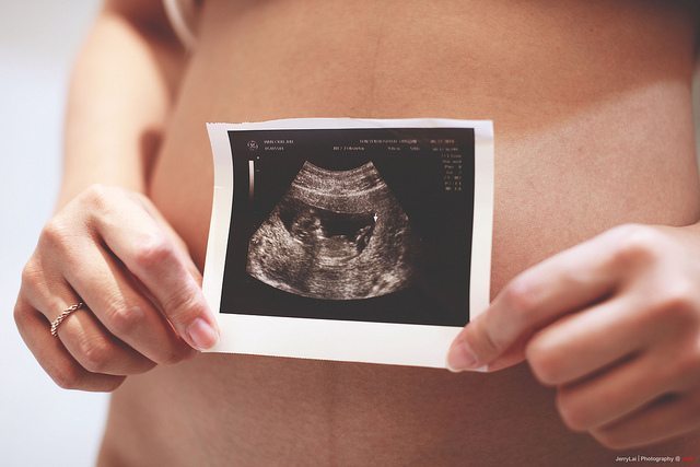
- Ultrasound: During the final trimester, doctors can take measurements of the baby’s different body parts in order to estimate weight.
- Abdominal circumference (AC) is the most reliable and common way to determine macrosomia risks. If two or more successive scans show an increase in the abdominal circumference, doctors can usually diagnose macrosomia.
- Leopold maneuver: A physician can estimate a baby’s weight by pushing on the mother’s abdomen.
- Fundal height measurements
How is Macrosomia Managed?
In pregnancies dealing with macrosomia, it is most important to manage the underlying causes of the condition. For instance, obese women should be instructed to gain as little weight as possible and see a dietician or nutritionist.
While vaginal delivery is still possible for macrosomic babies, the baby will risk certain injuries. Vaginal deliveries for macrosomic babies often require the use of vacuum extractors or forceps, and instrument delivery is a risk factor associated with a number of traumatic head injuries. Furthermore, the physician should be prepared to deal with shoulder dystocia, a situation in which the baby’s shoulders cannot pass through the birth canal. In all situations, the physician should be prepared to perform an emergency C-section. Most importantly, the physician should inform the patient of the risks of vaginally delivering a macrosomic baby.
A C-section delivery is recommended for most situations dealing with a macrosomic pregnancy. After week 39, physicians can take amniotic fluid samples to determine if the baby’s lungs are developed and may choose to perform the C-section thereafter.
Macrosomia and Medical Malpractice
It is crucial that physicians monitor both the mother and baby very closely when risk factors for macrosomia are present. Doctors and other medical professionals should be prepared to hold an emergency C-section or deliver the baby early, and in each case the patient should be informed of her situation, options, and potential delivery complications.
The following issues related to macrosomia deliveries may constitute negligence:
- The physician inappropriately used delivery assistance instruments like forceps or vacuum extractors .
- The physician failed to order a C-section.
- The physician caused a birth injury by forcing a vaginal birth.
- The physician failed to act promptly during an emergency, which in many cases may include the failure to quickly order and perform a C-section.
- A medical professional improperly prescribed or used labor-induction drugs like Pitocin or Cytotec.
- The physician failed to give the patient adequate informed consent and advice about the risks and delivery alternatives.
To find out if you have a case, contact our Michigan birth injury and macrosomia attorneys toll-free at (888) 592-1857. You may also Live Chat with our lawyers or fill out this online contact form. Our legal and medical teams understand the complex legal matters surrounding macrosomia cases and will assist you to obtain the information and compensation you deserve.
